


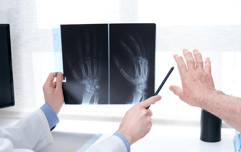
Arthrosis is a chronic disease of the joints associated with degenerative-dystrophic changes in their structures with predominant damage to cartilage tissue.The disease progresses slowly and is asymptomatic in the initial stages.The cartilage and bone tissue of the joint, its capsule and periarticular bursa are gradually destroyed, and later the surrounding muscles, ligaments and subcutaneous tissue are involved in the pathological process.
According to WHO, every tenth person suffers from this disease, and the risk of developing it increases significantly in people over 50 years of age.The knee and hip joints are most often affected.The cause of arthrosis is the inability of the joint to cope with the load on it.In response to traumatic effects, inflammation develops, which leads to pathological processes in the tissues of the joint.
Patients with arthrosis of the joints require complex treatment, without which the disease leads to decreased ability to work and quality of life, limitation of sports and professional activities and, ultimately, to disability.
Classification of arthrosis
Based on the cause of occurrence, the disease is divided into:
- primary arthrosis - occurs due to the fact that the cells of the cartilage tissue of the joint are destroyed faster than new ones are formed.Most often this is associated with age-related changes and is not a consequence of any specific pathological process in the body;
- secondary arthrosis - appears against the background of a specific disease or as a result of joint injury.
Based on location, arthrosis is divided into localized (affecting less than 3 joints) and generalized (affecting more than 3 joints).The latter is also called polyarthrosis.
Causes of arthrosis
This disease ranks first in the world among the causes of pain and impairment of activity.Despite the fact that age-related changes are considered the main cause, it is often diagnosed in young patients.Women are more prone to arthrosis than men, although closer to 70 years of age this difference becomes almost imperceptible.
Arthrosis of the hip, knee, ankle and shoulder joints has the greatest negative impact on the quality of life and ability to work of patients.Less common are arthrosis of the elbow and temporomandibular joints, as well as the hands, feet and spine.
Predisposing factors include:
- old age;
- genetic predisposition;
- intense physical activity associated with sports or professional activities, or, conversely, a sedentary lifestyle, which disrupts the normal nutrition of tissues, especially in large joints;
- joint injuries (fractures, cracks, dislocations);
- wearing inappropriate shoes;
- systemic diseases (metabolic disorders, endocrine diseases, neuropathies, gastrointestinal diseases, bleeding disorders);
- poor nutrition;
- overweight.
Women are more likely to suffer from this disease because they are more susceptible to hormonal imbalances.
Symptoms and degrees of arthrosis
The main clinical sign of arthrosis is pain.In the early stages it may be absent, mild or intermittent.As the disease progresses, the pain becomes more noticeable, it can occur during exercise, and in advanced stages it can be bothersome even at rest.
Secondary manifestations of the disease depend on the localization of the process.Most often, this is limited mobility in the affected area (especially after sleep or a long rest), deformation, swelling and redness in the joint area, crunching and crepitus in it when moving, sleep disturbance due to pain and the inability to find a comfortable body position, changes in gait and coordination, muscle spasms.
It is worth noting that the pathological process in one joint can move to neighboring ones due to improper load distribution or restrictions in movement.
According to the severity of the lesion, they are distinguished:
- arthrosis of the 1st degree - is asymptomatic or the signs are not expressed, the patient remains able to work, and diagnosis is difficult;
- Arthrosis of the 2nd degree – accompanied by severe symptoms, the patient’s quality of life decreases, diagnostics reveals obvious disorders;
- arthrosis of the 3rd degree - characterized by an increase in clinical signs, joint deformation is externally noted, and diagnostic methods reveal serious intra-articular disorders;
- arthrosis of the 4th degree - leads to partial or complete disability; disorders are found in all structures of the joint, surrounding muscles and ligaments.
Diagnosis of arthrosis
An orthopedic traumatologist makes a primary diagnosis based on the patient’s data on his lifestyle and work activities, the nature and duration of complaints, the dynamics of symptoms, the presence of arthrosis in relatives, and also conducts an examination and prescribes tests or consultations with related specialists (endocrinologist, hematologist, nutritionist, gastroenterologist).
Laboratory tests include a general blood test with a leukocyte count, hemoglobin level and ESR.These indicators indicate inflammation in the body, which is necessarily present with arthrosis.Biochemical parameters measure the level of rheumatoid factor and C-reactive protein.If certain pathologies are suspected, other laboratory blood parameters are checked.Also of diagnostic value is the analysis of synovial fluid, which reveals pathological cells and inclusions, allows you to confirm the diagnosis or differentiate another disease.
Instrumental and visual methods for diagnosing arthrosis help not only to detect pathology, but also to determine the stage of the disease and the degree of involvement of surrounding tissues.The most effective of them are:
- radiography - carried out mainly in two projections, it is used to detect narrowing of the joint space and the formation of bone growths (osteophytes) at the site of damaged cartilage;
- magnetic resonance and computed tomography - prescribed in the early stages, when minor changes are not yet noticeable on x-rays;
- Ultrasound - used in addition to these methods and helps to identify the accumulation of excess fluid in the joint cavity (for example, a Baker's cyst in gonarthrosis), assess the condition of the surrounding tissues, and measure the thickness of the articular cartilage;
- scintography – involves the intravenous administration of a radioactive drug that accumulates in tissues with inflammatory processes, and such areas of accumulation are displayed on the images, thanks to this it is possible to identify arthrosis in the early stages and carry out differential diagnosis;
- arthroscopy - makes it possible to examine the joint from the inside by introducing a microcamera into the articular cavity through a small incision, as a result, you can collect detailed data on the ongoing pathological processes and injuries, as well as take a biopsy from the affected area;
- histological examination of the synovial membrane - reveals an altered ratio of healthy cells and the presence of pathological inclusions in the joint, allowing for differential diagnosis.
Differential diagnosis allows you to distinguish arthritis from other diseases with a similar clinical picture, which include:
- various arthritis (ankylosing, reactive, rheumatoid, psoriatic, infectious);
- gout and pseudogout;
- diseases of muscles and ligaments (fibromyalgia, polymyalgia rheumatica);
- arthropathy (diabetic, paraneoplastic);
- congenital diseases (hypoplasia of the femoral head).
Treatment of arthrosis
Despite the widespread prevalence of the disease and the well-studied methods, there is no treatment for arthrosis.Therapeutic approaches are aimed at eliminating pain, relieving inflammation, restoring joint function and preventing complications.
The choice of therapy depends on the cause, location and degree of arthrosis:
- drug therapy in the form of taking tablets for arthrosis (painkillers and non-steroidal anti-inflammatory drugs, corticosteroids, chondroprotectors), intra-articular or periarticular injections of these groups of drugs, additionally using ointments and gels;
- physiotherapy during remission (medicinal electrophoresis, galvanization, acupuncture, electrical stimulation, shock wave therapy, massage, magnetic and laser therapy, cryotherapy);
- therapeutic exercises for arthrosis are prescribed individually, sessions are carried out under the supervision of a specialist;
- in advanced cases, surgical intervention is performed (partial or complete joint replacement, suturing or removal of a torn meniscus, removal of bone growths and Baker's cysts).
Prevention of arthrosis
It is recommended to control the load on the joints, maintain a healthy lifestyle, engage in therapeutic exercises, monitor weight and adhere to proper nutrition.
An orthopedic traumatologist will help select knee pads, an orthosis or a bandage for patients with arthrosis to fix the affected joint, reduce the load on it and prevent injury.
Preventive examinations and timely consultation with a doctor when discomfort in the joints occurs allows you to identify the problem at an early stage and avoid severe complications and disability.